Introduction to Human Anatomy
Conditions d’achèvement
1. Definition and Object of Human Anatomy
Anatomy is the branch of biological sciences that deals with the structure and organization of living organisms, particularly the human body.
The word anatomy comes from the Greek ana (“up”) and tome (“cutting”), meaning “to cut up” or “dissect.”
- Human anatomy focuses on the morphology, organization, and relationships between different organs and systems of the human body.
- The main objective of anatomy is to understand how the body is built and how its parts relate to each other in form and function.
- It serves as the foundation for all medical and biological sciences, especially physiology, pathology, and clinical medicine.
Branches of anatomy:
- Macroscopic (gross) anatomy: study of structures visible to the naked eye (e.g., bones, muscles, organs).
- Microscopic anatomy (histology): study of tissues and cells under a microscope.
- Developmental anatomy (embryology): study of structural changes from conception to adulthood.
- Functional anatomy: study of structures in relation to their functions.
- Topographical or regional anatomy: study of specific body regions (e.g., thorax, abdomen, limbs).
- Systemic anatomy: study of each organ system separately (skeletal, muscular, nervous, etc.).
2. Anatomical Position and Reference Planes
a. Anatomical Position
The standard anatomical position is a reference posture used to describe the body’s parts and their relations accurately.
In this position:
- The body stands upright.
- The head, eyes, and toes face forward.
- The upper limbs are placed along the sides of the body.
- The palms face forward (supinated position).
- The lower limbs are close together, with feet parallel and pointing forward.
This position allows clear and universal communication in anatomy.
b. Anatomical Reference Planes
Anatomical planes are imaginary lines used to describe cuts and directions in the body.
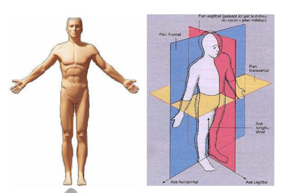
To accurately describe the location of structures and the directions of movements in the human body, anatomists use imaginary reference planes. These planes divide the body into specific sections and serve as a universal language to describe anatomical relationships. They are always defined with respect to the standard anatomical position—that is, the body standing upright, facing forward, with arms alongside the body and palms turned forward.
There are three principal anatomical planes: the median (or midsagittal) plane, the frontal (or coronal) plane, and the transverse (or horizontal) plane. Other additional or accessory planes may be drawn parallel to these main ones to describe regions more precisely.
a. The Median (Midsagittal) Plane
The median plane is a vertical plane that divides the body into two equal right and left halves. It passes through the midline of the body, running from the top of the head down through the center of the trunk to the feet. Structures located on or near this midline—such as the nose, mouth, sternum, umbilicus, and spinal column—are said to be medial.
Planes parallel to the median plane but situated on either side are called parasagittal planes. These divide the body into unequal right and left portions, allowing more specific anatomical descriptions. For instance, one can speak of a sagittal section through the shoulder or the thigh without necessarily being along the midline.
b. The Frontal (Coronal) Plane
The frontal plane, also known as the coronal plane, is another vertical plane, but it divides the body into anterior (front) and posterior (back) portions. This plane runs parallel to the forehead (from the Latin corona, meaning “crown”), extending vertically from one side of the body to the other.
Structures situated toward the front of this plane are described as anterior or ventral, while those toward the back are posterior or dorsal. The frontal plane is especially useful in clinical and imaging contexts, for example, in radiology when viewing coronal sections of the brain or thorax, where we can distinguish anterior structures like the sternum and ribs from posterior ones like the vertebral column.
c. The Transverse (Horizontal or Axial) Plane
The transverse plane, sometimes referred to as the horizontal plane or axial plane, divides the body into superior (upper) and inferior (lower) parts. It runs perpendicular to both the median and frontal planes, cutting the body horizontally at any chosen level—from the head down to the feet.
This plane is especially important in modern medical imaging (CT scans and MRI), where images are often presented as axial slices. Anatomists use this plane to describe relative positions such as above or below another structure. For example, the thoracic cavity is superior to the abdominal cavity, and the pelvis is inferior to the abdomen.
In dissection or imaging, when the body is “cut” along one of these planes, we obtain a section—such as a sagittal section, coronal section, or transverse section—allowing visualization of internal organization.
|
Plane |
Description |
Example |
|
Median (midsagittal) plane |
Divides the body into equal right and left halves. |
Nose and spine are on this plane. |
|
Sagittal plane |
Parallel to the median plane but not centered. |
Divides the body into unequal right and left parts. |
|
Frontal (coronal) plane |
Divides the body into anterior (front) and posterior (back) parts. |
Through the shoulders. |
|
Transverse (horizontal or axial) plane |
Divides the body into superior (upper) and inferior (lower) parts. |
Common in imaging (CT scans). |
3. Position of Anatomical Elements
To precisely locate body parts, several directional terms are used, always referring to the anatomical position:
- Superior (cranial): Toward the head or upper part of a structure. Example: The thorax is superior to the abdomen.
- Inferior (caudal): Toward the feet or lower part of a structure. Example: The stomach is inferior to the lungs.
- Anterior (ventral): Toward the front of the body. Example: The sternum is anterior to the heart.
- Posterior (dorsal): Toward the back of the body. Example: The spine is posterior to the sternum.
- Medial: Toward the midline of the body. Example: The big toe is medial to the little toe.
- Lateral: Away from the midline. Example: The thumb is lateral to the index finger.
- Proximal: Closer to the trunk or point of origin. Example: The shoulder is proximal to the elbow.
- Distal: Farther from the trunk or point of origin.Example: The hand is distal to the elbow.
- Superficial: Toward or on the surface. Example: The skin is superficial to the muscles.
- Deep: Away from the surface or more internal. Example: The bones are deep to the muscles.
These terms are fundamental for describing anatomical relationships and guiding medical imaging, surgery, and physical examination.
Table 1. Terms of Position and Direction of an Anatomical Element
|
Term |
Meaning |
Example |
|
Superior (cranial) |
Toward the head |
The heart is superior to the stomach. |
|
Inferior (caudal) |
Toward the feet |
The liver is inferior to the lungs. |
|
Anterior (ventral) |
Toward the front |
The sternum is anterior to the heart. |
|
Posterior (dorsal) |
Toward the back |
The spine is posterior to the heart. |
|
Medial |
Toward the midline |
The nose is medial to the eyes. |
|
Lateral |
Away from the midline |
The ears are lateral to the eyes. |
|
Proximal |
Nearer to the trunk or origin |
The elbow is proximal to the wrist. |
|
Distal |
Farther from the trunk or origin |
The fingers are distal to the wrist. |
|
Superficial |
Near the surface |
The skin is superficial to muscles. |
|
Deep |
Away from the surface |
The bones are deep to muscles. |
4. Movements of the Limbs
Movements are described relative to the anatomical position.
The human body is capable of a wide range of movements, most of which occur at the synovial joints, where bones articulate and muscles act to produce motion. These movements are always described with reference to the standard anatomical position, which serves as the universal point of orientation. Each movement occurs in a specific anatomical plane and around a corresponding axis.
In general, flexion and extension occur in the sagittal plane around a transverse axis, abduction and adduction occur in the frontal plane around an anteroposterior axis, and rotations occur in the transverse plane around a vertical axis. However, some complex movements such as circumduction combine multiple actions and planes simultaneously.
a. Flexion and Extension
Flexion refers to a movement that decreases the angle between two body segments or parts, bringing them closer together. It usually occurs when a body part moves forward from the anatomical position, except for the knee and toes where flexion moves posteriorly. For example, bending the elbow, bringing the forearm toward the arm, is flexion at the elbow joint. Similarly, bending the hip or shoulder to move the limb forward represents flexion.
In contrast, extension is the opposite movement — it increases the angle between body parts, moving them away from each other. For instance, straightening the elbow or knee returns the limb to its anatomical position, representing extension. When extension continues beyond the anatomical position, it is called hyperextension, as in bending the head backward or arching the spine.
b. Abduction and Adduction
Abduction means moving a body part away from the midline of the body or from the median plane. Raising the arm or leg laterally, away from the body, is an example of abduction. In the fingers and toes, abduction refers to movement away from the axis of the middle finger or the second toe.
Conversely, adduction denotes the movement of a limb or body part toward the midline or toward another part. Bringing the raised arm back to the side of the body is adduction of the shoulder joint. These two opposite movements are essential for coordinated motor control and balance, particularly in the upper and lower limbs.
c. Rotation (Medial and Lateral)
Rotation refers to movement around the longitudinal axis of a bone or body segment. When a limb rotates toward the median plane, it is called medial (internal) rotation. For example, turning the forearm so that the hand and fingers move inward toward the midline represents medial rotation of the shoulder.
On the other hand, lateral (external) rotation occurs when the limb turns away from the midline. An example is rotating the leg outward so that the toes point away from the other leg. These rotational movements are particularly important in joints such as the shoulder, hip, and vertebral column, where multidirectional mobility is necessary.
d. Circumduction
Circumduction is a complex, conical movement that combines four fundamental motions: flexion, extension, abduction, and adduction. The distal end of the limb moves in a circular path, while the proximal end remains relatively fixed. This movement is typically seen in ball-and-socket joints like the shoulder and hip. When performing circumduction, the limb describes a cone, and the tip of the cone corresponds to the point of rotation near the joint. This type of movement is essential in many sports and daily activities, such as swimming, throwing, or drawing large circles with the arm.
e. Pronation and Supination
These two movements are specific to the forearm and hand.
Supination occurs when the palm of the hand faces upward or forward, with the radius and ulna (the two bones of the forearm) lying parallel to each other — this is the position of the hand in the standard anatomical position.
Pronation is the opposite: the palm faces downward or backward, and the radius crosses over the ulna. A simple way to remember this is the phrase “you carry soup with supination,” since the hand holds a bowl in that position. These movements occur primarily at the proximal and distal radioulnar joints and are crucial for hand dexterity.
f. Inversion and Eversion
Inversion and eversion describe movements of the foot at the subtalar joint.
Inversion occurs when the sole of the foot turns inward, toward the median plane, as when you tilt the sole to face the other foot.
Eversion, on the contrary, is when the sole turns outward, away from the median plane. These movements play an essential role in balance and adaptation to uneven ground, though excessive inversion can result in ankle sprains.
g. Elevation and Depression
Elevation refers to moving a body part upward, while depression means moving it downward. For instance, when you shrug your shoulders, you are elevating the scapulae; when you relax them back down, you are performing depression. Similarly, elevation and depression occur with the jaw — lifting it to close the mouth (elevation) and lowering it to open the mouth (depression).
h. Opposition and Reposition
Specific to the thumb, opposition is the movement that allows the thumb to touch the tips of the other fingers — a unique and defining feature of the human hand. The reverse motion, moving the thumb back to its anatomical position, is called reposition. These two actions are fundamental to grasping and manipulating objects.
Summary
Movements of the limbs are fundamental expressions of human anatomy and function. Each motion is defined according to the anatomical planes and axes, allowing precise description of how the body interacts with its environment. Understanding these movements provides the basis for studying biomechanics, muscle function, and joint physiology, all essential components of anatomy and human movement sciences.
|
Type of Movement |
Definition |
Example |
|
Flexion |
Decreasing the angle between two parts |
Bending the elbow |
|
Extension |
Increasing the angle between two parts |
Straightening the elbow |
|
Abduction |
Moving away from the midline |
Raising the arm sideways |
|
Adduction |
Moving toward the midline |
Lowering the arm to the side |
|
Rotation |
Turning around a longitudinal axis |
Turning the head left or right |
|
Medial (internal) rotation |
Rotation toward the midline |
Turning the leg inward |
|
Lateral (external) rotation |
Rotation away from the midline |
Turning the leg outward |
|
Circumduction |
Circular movement combining flexion, extension, abduction, and adduction |
Making a circle with the arm |
|
Pronation |
Turning the forearm so the palm faces down |
Palm down |
|
Supination |
Turning the forearm so the palm faces up |
Palm up |
|
Eversion |
Turning the sole of the foot outward |
Sole outward |
|
Inversion |
Turning the sole of the foot inward |
Sole inward |
|
Elevation |
Lifting a body part upward |
Shrugging shoulders |
|
Depression |
Moving a body part downward |
Lowering shoulders |
5. Introduction to Osteology, Arthrology, and Myology
a. Osteology (Study of Bones)
- Osteology is the branch of anatomy that studies the bones of the human skeleton.
- The human body contains 206 bones in the adult.
- Functions:
- Support of the body.
- Protection of vital organs.
- Movement (as levers for muscles).
- Mineral storage (especially calcium and phosphorus).
- Hematopoiesis (production of blood cells in bone marrow).
Classification of bones:
- Long bones (e.g., femur, humerus) – found in limbs.
- Short bones (e.g., carpal bones) – provide stability.
- Flat bones (e.g., skull, sternum) – protect organs.
- Irregular bones (e.g., vertebrae).
- Sesamoid bones (e.g., patella) – embedded in tendons.

b. Arthrology (Study of Joints)
- Arthrology is the study of joints or articulations, the connections between bones.
- Functions: allow movement, provide stability, and connect skeletal elements.
Classification of joints:
- Fibrous joints – no movement (e.g., sutures of the skull).
- Cartilaginous joints – limited movement (e.g., intervertebral discs).
- Synovial joints – freely movable (e.g., shoulder, knee, hip).
|
Type of Joint |
Description |
Example in Upper Limb |
|
Fibrous joints |
Bones united by fibrous tissue, minimal movement |
Interosseous membrane between radius and ulna |
|
Cartilaginous joints |
Bones connected by cartilage |
Sternoclavicular joint (partly) |
|
Synovial joints |
Joint cavity with synovial fluid, allowing free movement |
Most upper limb joints (shoulder, elbow, wrist, etc.) |
Components of a synovial joint:
- Articular surfaces covered with cartilage.
- Joint capsule and synovial membrane.
- Synovial fluid for lubrication.
- Ligaments and muscles for stability.
c. Myology (Study of Muscles)
- Myology is the branch of anatomy dealing with muscles and their structures, types, and functions.
Functions of muscles:
- Produce movement.
- Maintain posture.
- Stabilize joints.
- Generate heat.
Classification of muscles:
- Skeletal (striated voluntary) – attached to bones, controlled voluntarily.
- Cardiac (striated involuntary) – found in the heart.
- Smooth (non-striated involuntary) – found in walls of viscera (stomach, vessels).
Each skeletal muscle has:
- Origin (fixed point of attachment)
- Insertion (movable attachment)
- Action (type of movement produced)
- Innervation (nerve supply)
6. Summary
Anatomy provides the structural foundation for understanding human function.
- It defines how the body is organized into systems and regions.
- Knowledge of anatomical positions, planes, and movements is essential for describing any part precisely.
- Osteology, arthrology, and myology together explain how the body supports, connects, and moves.

Modifié le: mercredi 4 février 2026, 20:51